


Tutorial
These icons indicate there is something to be interacted with. Click it when you see it.
Tutorial
These icons indicate there is something to be interacted with. Click it when you see it.
Tutorial
These icons indicate there is something to be interacted with. Click it when you see it.
touchIN CONVERSATION
 A relaxed discussion between two faculty focussed on real world clinical issues. Useful tips below will show how to navigate the activity. Join the conversation.
Close
A relaxed discussion between two faculty focussed on real world clinical issues. Useful tips below will show how to navigate the activity. Join the conversation.
Close
A relaxed discussion between two faculty focussed on real world clinical issues. Useful tips below will show how to navigate the activity. Join the conversation.
Close
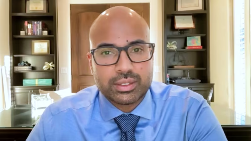
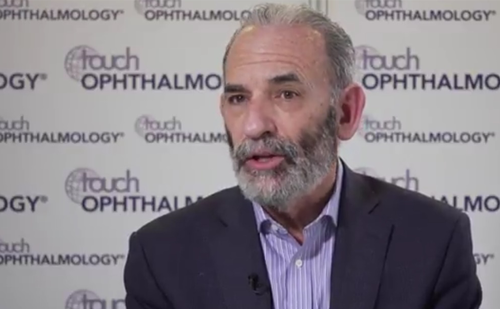
Recent advances in the care of patients with geographic atrophy: Bringing new and emerging therapies into focus
Learning Objectives
After watching this activity, participants should be better able to:
- Outline the progression of geographic atrophy (GA) and approaches to support early diagnosis and intervention
- Interpret the latest data on mechanisms of action, efficacy and safety for agents used in the treatment of GA and their implications for clinical practice
- Discuss the value of imaging techniques, metrics and endpoints for the treatment of GA and how they may be incorporated into clinical practice
Overview
In this activity, two experts discuss recent advances in the care of patients with geographic atrophy (GA). They explore the importance of early identification of GA and how this can be achieved, examine the potential impact of new and emerging therapies for GA, and highlight key practical considerations for monitoring treatment outcomes and evaluating effectiveness of therapy in clinical practice.
Target Audience
This activity has been designed to meet the educational needs of ophthalmologists, retina specialists and optometrists involved in the management of patients with GA.
USF Accreditation
Disclosures
USF Health adheres to the Standards for Integrity and Independence in Accredited Continuing Education. All individuals in a position to influence content have disclosed to USF Health any financial relationship with an ineligible organization. USF Health has reviewed and mitigated all relevant financial relationships related to the content of the activity. The relevant relationships are listed below. All individuals not listed have no relevant financial relationships.
Faculty
Prof. Giuseppe Querques discloses: Consultant and/or advisory board fees from 4DMT, AbbVie, Apellis, Bausch & Lomb, Bayer, Boehringer Ingelheim, EyePoint, Heidelberg Engineering, iCARE-CenterVue, Lumithera, Novartis, Roche, Sandoz, Théa Pharmaceuticals and Zeiss.
Dr Arshad M. Khanani discloses: Consultant fees from 4DMT, AbbVie, ADARx Pharmaceuticals, Adverum, Alcon, Alkeus, Allgenesis, Amgen, Annexin, Annexon, Apellis Pharmaceuticals, Ashvattha Therapeutics, Astellas, Aviceda Therapeutics, Beacon Therapeutics, Boehringer Ingelheim, Clearside Biomedical, Complement Therapeutics, Exegenesis, EyePoint Pharmaceuticals, Fronterra Therapeutics, Genentech, Gyroscope Therapeutics, Harrow, i-Lumen Scientific, InFocus, Iveric Bio, Janssen Pharmaceuticals, Kodiak Sciences, Kriya Therapeutics, Kyowa Kirin, Merit, Neurotech, Nanoscope, Novartis, Ocular Therapeutix, Oculis, Ocuphire, OcuTerra, Ollin, Olive BioPharma, Opthea, Opus Genetics, Oxular, Oxurion, Perfuse, Ray Therapeutics, Recens Medical, Regeneron Pharmaceuticals, Regenxbio, Revive, RevOpsis, Roche, Samsung Biologics, Sanofi, Stealth BioTherapeutics, Surrozen, Théa Pharmaceuticals, Therini, Unity Biotechnology, Vanotech, Vial and ZipBio. Grants/research support from 4DMT, Adverum, Alexion, Annexon, Apellis Pharmaceuticals, Astellas, Aviceda Therapeutics, Complement Therapeutics, Eyepoint Pharmaceuticals, Exegenesis, Genentech, Gyroscope Therapeutics, Iveric Bio, Janssen, Kodiak, Kyowa Kirin, Neurotech, Ocular Therapeutix, Oxular, Regenxbio, Roche, Sanofi and Vanotech. Stock/Shareholder (self-managed) Ashvattha Therapeutics, Aviceda Therapeutics, Oculis, Ollin, Opthea, PolyPhotonix, Recens Medical, Perfuse, RevOpsis, Vial and ZipBio.
Content Reviewer
Danielle Walker, DNP, APRN, AGNP-C, has no financial interests/relationships or affiliations in relation to this activity.
Touch Medical Contributor
Hannah Fisher has no financial interests/relationships or affiliations in relation to this activity.
USF Health Office of Continuing Professional Development and touchIME staff have no financial interests/relationships or affiliations in relation to this activity.
Requirements for Successful Completion
In order to receive credit for this activity, participants must review the content and complete the post-test and evaluation form. Statements of credit are awarded upon successful completion of the post-test and evaluation form.
If you have questions regarding credit please contact cpdsupport@usf.edu
Accreditations
Physicians
This activity has been planned and implemented in accordance with the accreditation requirements and policies of the Accreditation Council for Continuing Medical Education (ACCME) through a joint providership of USF Health and touchIME. USF Health is accredited by the ACCME to provide continuing medical education for physicians.
USF Health designates this enduring material for a maximum of 0.75 AMA PRA Category 1 CreditsTM. Physicians should claim only the credit commensurate with the extent of their participation in the activity.
Advanced Practice Providers
Physician Assistants may claim a maximum of 0.75 Category 1 credits for completing this activity. NCCPA accepts AMA PRA Category 1 CreditTM from organizations accredited by ACCME or a recognized state medical society.
The AANPCP accepts certificates of participation for educational activities approved for AMA PRA Category 1 CreditTM by ACCME-accredited providers. APRNs who participate will receive a certificate of completion commensurate with the extent of their participation.
Date of original release: 8 May 2025. Date credits expire: 8 May 2026.
If you have any questions regarding credit, please contact cpdsupport@usf.edu
EBAC® Accreditation
touchIME is an EBAC® accredited provider since 2023.
This program is accredited by the European Board for Accreditation of Continuing Education for Health Professionals (EBAC®) for 50 minutes of effective education time.
EBAC® holds an agreement on mutual recognition of substantive equivalency with the US Accreditation Council for CME (ACCME) and the Royal College of Physicians and Surgeons of Canada, respectively.
Through an agreement between the European Board for Accreditation of Continuing Education for Health Professionals (EBAC®) and the American Medical Association, physicians may convert EBAC® External CME credits to AMA PRA Category 1 Credits™. Information on the process to convert EBAC® credit to AMA credit can be found on the AMA website. Other healthcare professionals may obtain from the AMA a certificate of having participated in an activity eligible for conversion of credit to AMA PRA Category 1 Credit™.
EBAC® is a member of the International Academy for CPD Accreditation (IACPDA) and a partner member of the International Association of Medical Regulatory Authorities (IAMRA).
Faculty Disclosure Statement/Conflict of Interest Policy
In compliance with EBAC® guidelines, all speakers/chairpersons participating in this programme have disclosed or indicated potential conflicts of interest which might cause a bias in the presentations. The Organizing Committee/Course Director is responsible for ensuring that all potential conflicts of interest relevant to the event have been mitigated and declared to the audience prior to the CME activities.
Requirements for Successful Completion
Certificates of Completion may be awarded upon successful completion of the post-test and evaluation form. If you have completed one hour or more of effective education through EBAC® accredited CE activities, please contact us at accreditation@touchime.org to receive your EBAC® CE credit certificate. EBAC® grants 1 CE credit for every hour of education completed.
Date of original release: 8 May 2025. Date credits expire: 8 May 2027.
Time to complete: 50 minutes
If you have any questions regarding the EBAC® credits, please contact accreditation@touchime.org
To obtain the CE/CME credit(s) from this activity, please complete this post-activity test.
Claim CreditYou may also be interested in...

touchIN CONVERSATION
Recent advances in the care of patients with geographic atrophy: Bringing new and emerging therapies into focus
This activity is CE/CME accredited
CONFERENCE HUB
Jordana Fein, ARVO 2023: Phase II CANDELA study post hoc analysis: intravitreal aflibercept injection 8 mg versus 2 mg

touchCONGRESS
Will a scientific breakthrough translate into clinical benefits in nAMD? Highlights from EURETINA 2019
CONFERENCE HUB
Baruch Kuppermann, ISOPT 2018 – Challenges in the treatment of dry AMD

JOURNALS
Effectiveness of the Dexamethasone Intravitreal Implant for Treatment of Patients with Diabetic Macular Oedema
JOURNALS
Diabetic Retinopathy Treatment

REGISTER NOW FOR FREE ACCESS TO
- 1000+ topical and insightful peer-reviewed journal articles
- 100+ hours of bite-sized congress highlights
- 10 major therapy areas packed with the latest scientific advances
- 150+ specialties offering learn-on-the-go medical education
- + Concise email updates and newsletters so you never miss out

Log into your Touch Account
Earn and track your CME credits on the go, save articles for later, and follow the latest congress coverage.
Sign up with an Email
Or use a .
This Functionality is for
Members Only
Explore the latest in medical education and stay current in your field. Create a free account to track your learning.
